Shrimp in Group A are infected with EHP, exhibiting greater size variation and signs of hepatopancreas atrophy (Nkuba et al., 2021)
Understanding EHP and Its Symptoms
Enterocytozoon hepatopenaei (EHP) is a parasitic disease that has had a profound impact on the farming of white shrimp (Litopenaeus vannamei) and black tiger shrimp (Penaeus monodon). It is an intracellular parasite that infects the epithelial cells of the shrimp's hepatopancreatic tubules. Unlike other diseases such as WSSV or AHPND, EHP does not directly cause mortality. However, it significantly impairs shrimp growth, leading to stunted development and reduced yields.
Common symptoms of EHP infection include:
- Slow Growth: Infected shrimp exhibit a noticeably slower growth rate compared to healthy shrimp.
- Uneven Sizes: Shrimp within the same batch display significant size variations.
- Poor FCR: Despite regular feeding, the shrimp’s growth remains suboptimal.
- Abnormal Hepatopancreas Appearance: The hepatopancreas may show discoloration or morphological abnormalities.
- No Noticeable Mortality Spike: Unlike other diseases, EHP typically does not cause sudden mass mortality, making early detection more challenging.
However, these symptoms are not exclusive to EHP and may also result from other environmental or nutritional factors. Therefore, using scientific methods for supplementary diagnosis is crucial. Below is a brief introduction to the main diagnostic methods currently used for EHP.
Diagnostic Methods for EHP
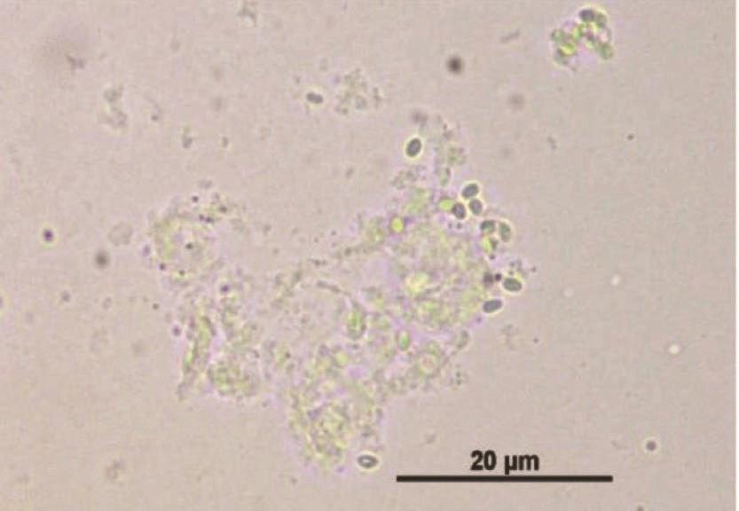
1. Microscopic Examination Microscopic examination is one of the simplest methods to assess shrimp health and identify EHP infection by directly examining feces, hepatopancreas, and intestinal tissues. However, the complexity of substances in the digestive tract and feces can make interpretation difficult.
Steps for Microscopic Detection:
- Collect feces, hepatopancreas, and intestinal tissue samples from suspected shrimp.
- Place the sample directly on a glass slide and cover it with a coverslip, or mix the sample with saline solution before covering it with a coverslip and gently pressing to create a smear sample.
- Observe the hepatopancreas' health under a compound microscope at 40x magnification.
- Observe at 400x magnification for the possibility of detecting EHP spores.
- EHP spores are typically oval or ovoid, measuring approximately 1.1–1.4 µm.
Advantages:
- Cost-effective and quick.
- Suitable for preliminary diagnosis at the farm level.
Disadvantages:
- Requires trained personnel to accurately identify EHP spores.
- The abundance of feed particles and large organic molecules in the digestive tract and feces can obscure the slide view, making it challenging to observe EHP spores accurately.
- If the infection level is low, the number of EHP spores in the tissue may be too small to detect.
EHP spore under microscope(Praveena et al., 2018)
2. PCR (Polymerase Chain Reaction) PCR is considered the gold standard for diagnosing EHP infection due to its high sensitivity and specificity. This method can accurately detect trace amounts of EHP DNA in infected shrimp tissue or water samples from farming ponds.
Steps for PCR Detection:
- Extract DNA from hepatopancreas or fecal samples.
- Use EHP-specific primers to amplify the target DNA.
- Run the PCR product on an agarose gel and visualize the results under UV light.
Advantages:
- High sensitivity and specificity.
- Can detect even early-stage, mild infections.
Disadvantages:
- Requires specialized equipment and skilled technicians. Although portable PCR devices are now available for commercial use, operational technical requirements remain high.
- High detection costs, which may be unaffordable for many farmers.
How EHP Spreads Among Shrimp
To effectively implement biosecurity measures against EHP, it is crucial to understand how EHP spreads among shrimp.
Transmission Pathways of EHP
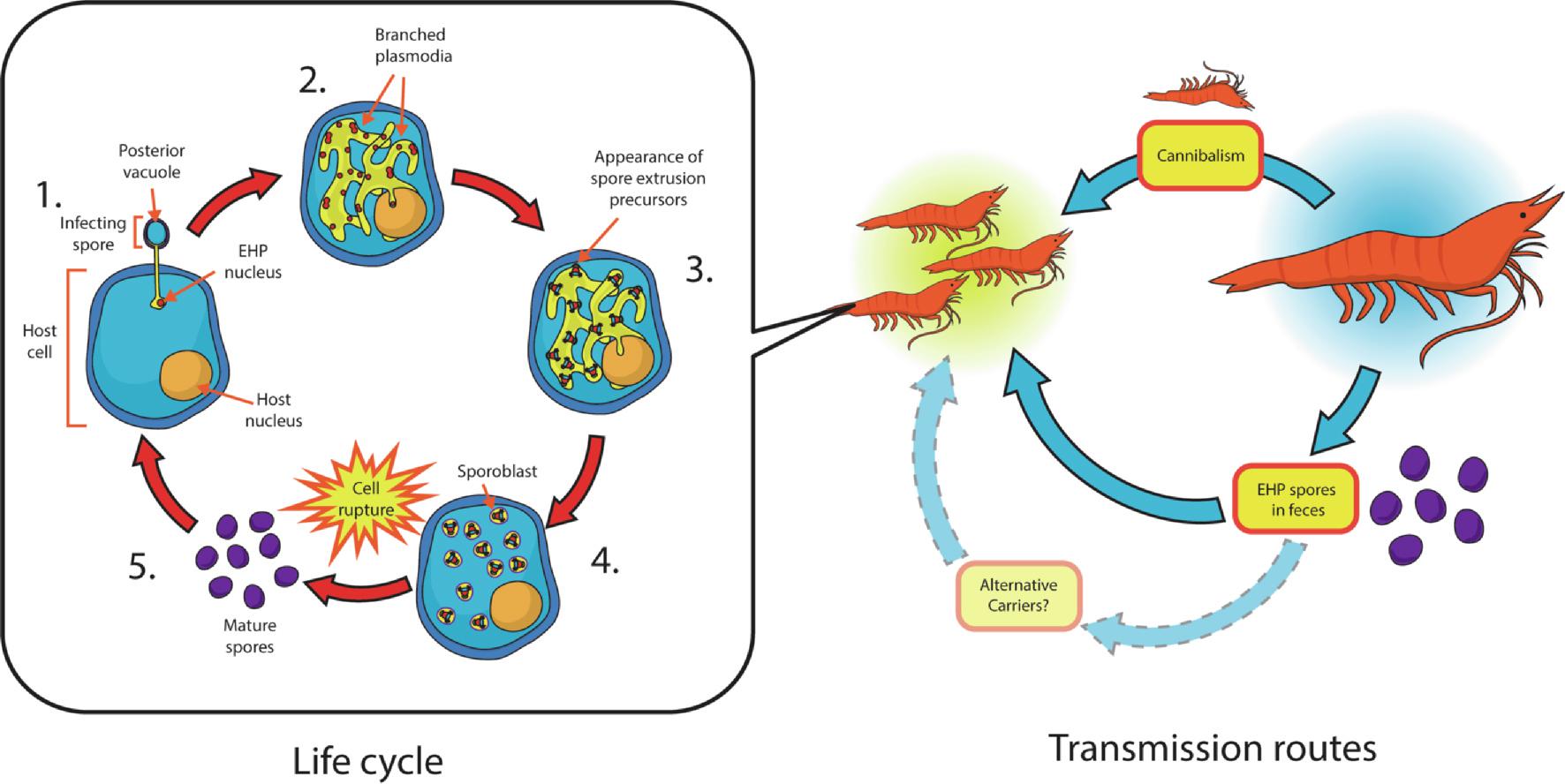
EHP is primarily transmitted through horizontal transmission.
Free-floating EHP spores in the environment are ingested by healthy shrimp. Once inside the shrimp, EHP begins its lifecycle in the intercellular stage. At this stage, EHP spores use a polar tube to infect shrimp hepatopancreas cells. The spores then proliferate inside the cells, producing a large number of new spores, which are eventually excreted into the environment through the shrimp’s feces.
Healthy shrimp ingest EHP spores by consuming infected shrimp carcasses or feces, leading to the spread of infection throughout the pond.
EHP life cycle(Chaijarasphong et al., 2021)
Post-Diagnosis Management Measures
Understanding the transmission mechanism of EHP helps guide effective management strategies. Once EHP infection is diagnosed in a pond, the focus shifts from prevention to control and management. In addition to strengthening biosecurity to prevent the infection from spreading to other ponds, several key measures should be implemented:
1. Improving Water Quality
Increase water exchange, reduce ammonia and nitrite levels, maintain dissolved oxygen levels and stable water conditions, minimize environmental stress on shrimp.
2. Adding Probiotics
Proper use of probiotics can help stabilize water quality, enhance shrimp immunity, and suppress potential pathogens. Some probiotics can inhibit Vibrio and other secondary infections, and even reduce EHP infection levels.
3. Using High-Quality Feed Additives
The main symptoms of EHP infection include slow growth and weakened immunity. Using natural extract-based feed additives, such as BiomiXin’s EHPurge, can boost shrimp growth and immunity, helping to reduce EHP infection while shortening the culture period and lowering the risk of various diseases.
Author: Barry Cheng, Manager of Aquaculture Department
Reference: Nkuba et al., 2021; Praveena et al., 2018; Chaijarasphong et al., 2021.